Research Makes Front Cover
Multiple Infusion Start Time Mass Spectrometry Imaging of Dynamic SIL-Glutathione Biosynthesis Using Infrared Matrix-Assisted Laser Desorption Electrospray Ionization
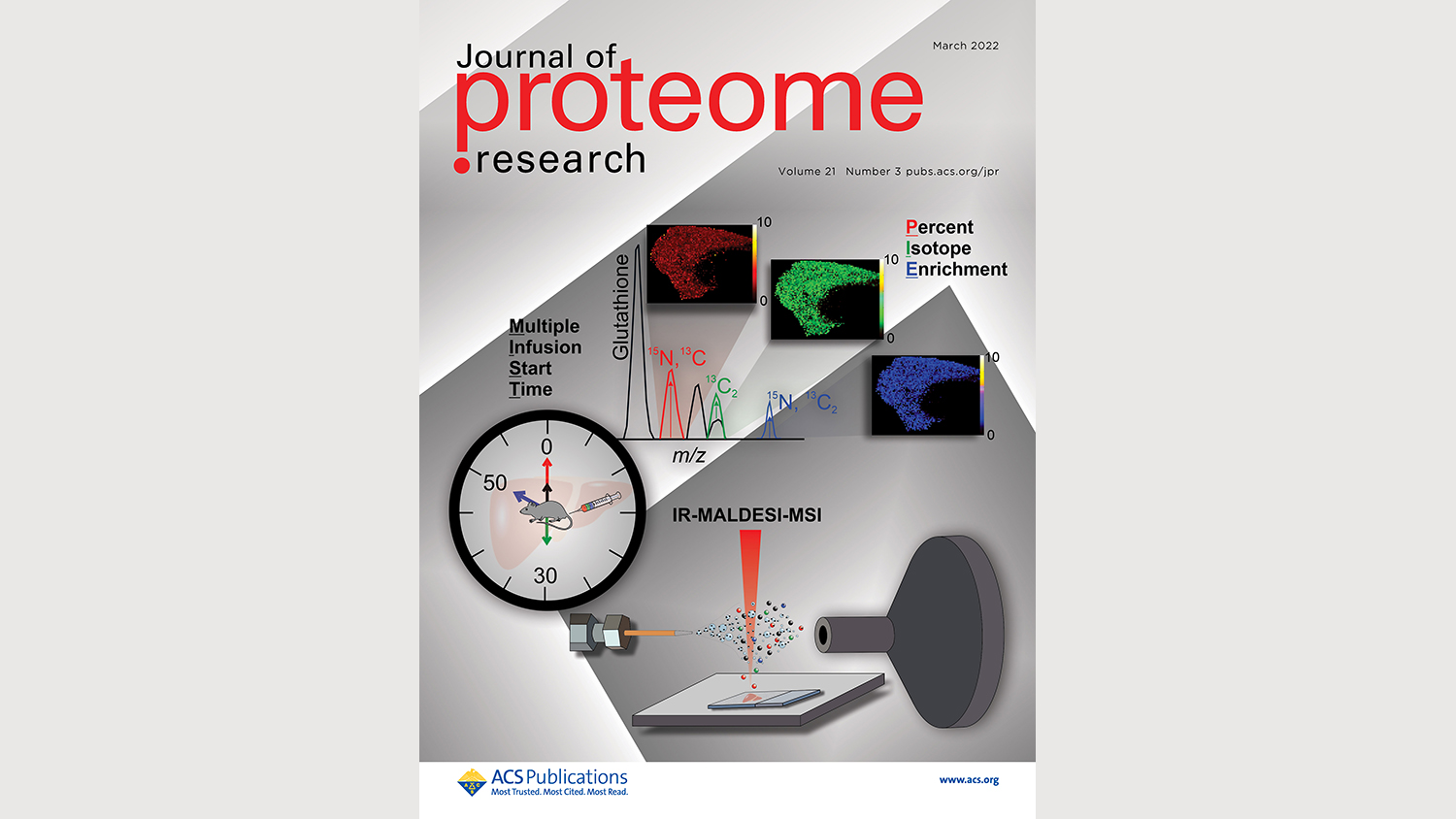
Innovative research at NC State makes one step further toward the global fight against cancer.
NC State researchers within the Department of Chemistry, in collaboration with the Department of Biomedical Engineering, published a recent discovery of significance to cancer research. The article, prepared by Allyson Mellinger, Kenneth Garrard, Sitora Khodjaniyazova, Zahid Rabbani, Michael Gamcsik, and David Muddiman was accepted for publication in the Journal of Proteome Research‘s special issue covering Metabolomics research of March 4, 2022. The cover artwork was also selected for print in the issue.
In this work, Mellinger et al. develop a novel method to measure and visualize the biosynthesis of the important antioxidant glutathione in mouse tissues.
To do so, stable isotope labeling and mass spectrometry imaging are leveraged via infrared matrix-assisted laser desorption electrospray ionization (IR-MALDESI).
What is novel about this method is the use of a multiple infusion start time (MIST) protocol for labeling the glutathione molecules, allowing three different time points to be measured in a single slice of tissue.
The method is projected to be used in the study of changes in glutathione biosynthesis across various types of cancer tissue models in order to learn more about how this pathway is involved in tumor progression.
Check out the article for further information on the matter.